LatticeZero: Physics-Based Molecular Scoring with WebGPU Acceleration
LatticeZero Technical White Paper v0.9.7 - February 2026
LatticeZero Team
Abstract
LatticeZero is a browser-based molecular docking and scoring platform that combines a 14-term physics-based energy function with per-target weight optimization and WebGPU-accelerated compute. We validate performance across 27 DEKOIS2 targets using tie-corrected 5-fold cross-validation, Y-scramble null controls, and holdout validation. On a 22-target overlap with published Glide SP benchmarks, LatticeZero IsoPose achieves a mean ROC-AUC of 0.821 versus 0.741 for Glide SP, winning 16 of 22 matchups (73%). The platform delivers IsoScore rescoring at 4,000+ ligands/second and IsoPose docking at ~3 seconds/ligand, all executing client-side via WebGPU compute shaders with zero server-side compute. We report all 27 target results including 6 targets below 0.60 AUC, one Y-scramble failure (COX2), and honest disclosure of significant AUC drops following our tie-handling bug correction. A REST API (v1) provides programmatic access.
1. Introduction
Molecular docking remains a cornerstone of structure-based drug discovery, yet practitioners face a persistent tension between physical rigor and practical throughput. Traditional physics-based scoring functions (GOLD, Glide XP) evaluate interactions from first principles but require expensive computational infrastructure and per-seat licensing. Machine learning approaches achieve speed but sacrifice interpretability - a neural network score of -7.3 offers no insight into which interactions drive binding.
LatticeZero addresses this gap with three design principles:
-
Physics from first principles. Every scoring term maps to a measurable physical interaction (dispersion, electrostatics, hydrogen bonding, desolvation). No training on binding affinity databases. No learned potentials. When a compound scores well, a medicinal chemist can inspect why - "strong electrostatic complementarity with Glu384" rather than "high predicted pIC50."
-
Target-specific weight profiles. Raw physics terms have different relative importance depending on the binding site. A zinc metalloprotease rewards metal coordination; a kinase rewards hinge hydrogen bonds. LatticeZero maintains a library of validated scoring profiles, each assigning optimized weights to the 14 physics terms for a specific target or target class.
-
Browser-native execution. All scoring computation runs client-side via WebGPU compute shaders. The user's GPU performs the molecular physics calculations. No compound structures are transmitted to any server. This architecture delivers both performance (4,000+ ligands/second for rescoring) and data privacy (structures never leave the client device).
This paper describes the scoring function architecture, validation methodology, and benchmark results as of version 0.9.7 (February 2026). We report all results transparently, including failures and limitations.
2. Methods
2.1 Scoring Function Architecture
LatticeZero uses a grid-based scoring approach. During target preparation, a 3D grid of pre-computed interaction energies is compiled for the binding pocket at 1.0 A spacing with 8.0 A padding. At scoring time, each ligand atom's position is mapped to the grid and its contributions are evaluated in parallel using WebGPU compute shaders.
The total score is a weighted linear combination of 14 physics-based energy terms:
Score = sum(w_i * E_i) for i = 1..14
More negative scores indicate stronger predicted binding. The 14 weights (w_i) constitute the scoring profile for a given target.
2.2 Physics Energy Terms
The scoring function comprises 14 terms in 8 categories:
| # | Term | Category | Physical Basis | Sign Convention |
|---|---|---|---|---|
| 1 | E_disp | Van der Waals | London dispersion (r^-6 attractive) | Negative = favorable |
| 2 | E_rep | Van der Waals | Pauli repulsion (r^-12 repulsive) | Positive = unfavorable |
| 3 | E_coul | Electrostatics | Coulombic charge-charge with distance-dependent dielectric | Negative = favorable |
| 4 | E_hbond | Hydrogen bonding | Directional D-H...A potential (distance + angle) | Negative = favorable |
| 5 | E_desolv | Desolvation | Atomic solvation parameter x buried surface area | Positive = penalty |
| 6 | E_clash | Steric | Close-contact penalty (hard overlap) | Positive = unfavorable |
| 7 | burial | Shape | Fraction of ligand surface buried by receptor | Negative = favorable |
| 8 | depth | Shape | Ligand penetration into pocket interior | Negative = favorable |
| 9 | strain | Conformational | Internal ligand torsional strain | Positive = unfavorable |
| 10 | aromaticBurial | Aromatic | Aromatic ring surface buried by receptor | Negative = favorable |
| 11 | hbondGeo | H-bond quality | Geometric quality of H-bond angle/distance | Negative = favorable |
| 12 | E_aromatic | Aromatic | Pi-stacking + cation-pi + aromatic interactions | Negative = favorable |
| 13 | contactArea | Shape | Ligand-receptor contact surface area | Negative = favorable |
| 14 | E_metal | Metal coordination | Metal ion coordination energy (Zn, Mg, Fe, etc.) | Negative = favorable |
GPU vs CLI implementation. Of the 14 terms, the following are computed in the WebGPU path: E_disp, E_rep, E_coul, E_hbond, E_clash, burial, and aromaticBurial. The remaining terms (E_desolv, depth, strain, hbondGeo, E_aromatic, contactArea, E_metal) are available in the CLI backend only and are set to zero in browser-only scoring runs. All benchmark results in this paper were generated using the CLI backend with all 14 terms active.
Parameter provenance. Van der Waals radii follow Bondi (1964) and Rowland & Taylor (1996): C 1.70, N 1.55, O 1.52, S 1.80, H 1.20 A, with metal-specific radii (Zn 1.39, Mg 1.73 A). Partial charges are computed via RDKit Gasteiger with element-heuristic fallback. Hydrogen bond parameters: max D-H...A distance 3.0 A, optimal 2.5 A, minimum angle 120 degrees, energy scaled by k_hb = -2.5 kcal/mol. Electrostatics use a distance-dependent dielectric (eps = 4.0 at contact to 40.0 at long range) with 8.0 A cutoff.
No external force-field parameter files (OPLS, AMBER) are loaded. All constants are versioned in code.
Functional Forms (GPU-Implemented Terms)
The core physics terms implemented in the WebGPU compute shader use the following functional forms. For each ligand-receptor atom pair at distance r with combined van der Waals diameter sigma = r_i + r_j:
Dispersion (London attraction):
E_disp = -0.5 * sum_pairs( (sigma/r)^6 )
Repulsion (Pauli exclusion):
E_rep = 0.1 * s_R * sum_pairs( (sigma/r)^12 )
where s_R is a softening parameter (0 < s_R <= 1.0; default 1.0). A ratio cap prevents numerical overflow at close contacts. The combined form is a scaled Lennard-Jones 12-6 potential with separate weights for the attractive and repulsive branches.
Coulomb (electrostatics with distance-dependent screening):
E_coul = k * sum_pairs( q_i * q_j / (eps(r) * r_eff) )
where q_i, q_j are Gasteiger partial charges (Gasteiger & Marsili, 1980), k is the Coulomb constant, r_eff = sqrt(r^2 + delta^2) is a damped distance (delta = 0.5 A) to avoid singularity, and the dielectric function is:
eps(r) = eps_min + (eps_max - eps_min) * (1 - exp(-r / lambda))
with eps_min = 4.0 (protein interior), eps_max = 40.0 (bulk water), and lambda = 4.0 A (transition length). This models the transition from low-dielectric protein interior at short range to high-dielectric solvent-like screening at long range.
Hydrogen bonding:
E_hbond = k_hb * f(theta) * g(r)
where k_hb = -2.5 kcal/mol, f(theta) is an angular dependence on the D-H...A angle (minimum 120 degrees), and g(r) is a distance function with optimal distance 2.5 A and maximum 3.0 A.
Steric clash:
E_clash = sum_pairs( max(0, 1 - r / ((r_i + r_j) * f_clash)) )
where f_clash is the clash factor controlling sensitivity. Each pair contributes a linear penalty proportional to the fractional overlap beyond the scaled VDW contact distance.
Burial (shape complementarity):
burial = (1/N_lig) * sum_pairs( 1 - r/r_cutoff ) for r < r_cutoff
where N_lig is the ligand heavy atom count and r_cutoff is the burial cutoff distance. This measures the fraction of ligand surface enclosed by receptor atoms.
2.3 Geometric Features (IsoPose)
In addition to the 14 physics terms, IsoPose evaluates 6 geometric features of each docked pose, for a total of 20 scoring features:
| Feature | Description |
|---|---|
| insideFrac | Fraction of ligand heavy atoms inside the defined pocket volume |
| triadScore | Quality of key pharmacophoric interactions (H-bond triads) |
| aromaticBurial | Fraction of aromatic ring surface buried by receptor |
| clashCount | Number of severe ligand-receptor atom overlaps |
| keyguideScore | Alignment with pharmacophore guide points (KeyGuide system) |
| waterAnchorFrac | Fraction of conserved water-mediated contacts preserved |
The KeyGuide system defines per-target pharmacophore anchor points (H-bond donors/acceptors, hydrophobic hotspots) and measures how well each pose satisfies them. KeyGuide features are configurable per target profile.
2.4 Pose Quality Score (PQS)
Every scored pose receives a Pose Quality Score classification:
| Verdict | Criteria |
|---|---|
| GOOD | Low clashes, ligand inside pocket, acceptable strain |
| FIXABLE | Moderate clashes or partial pocket occupancy |
| TRASH | Severe clashes, ligand outside pocket, or extreme strain |
PQS serves as a quality gate: even poses with favorable total scores are flagged if physically implausible. The distribution of PQS verdicts across a screening run is a useful diagnostic for input quality.
2.5 Pose Generation (IsoPose)
IsoPose performs de novo pose generation using a genetic algorithm (GA) with torsional sampling:
- Conformer generation - RDKit ETKDG generates initial 3D coordinates from SMILES
- GA search - Translational, rotational, and torsional degrees of freedom are evolved over multiple generations with configurable seeds, population size, and evaluation budget
- Scoring - Each pose is evaluated against the pre-computed interaction grid using 20 features (14 physics + 6 geometric)
- Selection - Top-K poses are retained per ligand, ranked by weighted score
Default parameters: 2 GA seeds, top-5 poses, 5000 max evaluations. These can be adjusted per-target via the profile system.
2.6 Target-Specific Scoring Profiles
Raw physics terms have different magnitudes and relative importance across target classes. A scoring profile assigns weights to each of the 14 physics terms (and optionally the 6 geometric features), tuning the function for a specific target:
- Metalloenzymes (ACE) upweight E_metal and E_coul for metal coordination and electrostatic complementarity
- Kinases (CDK2, EGFR) emphasize E_hbond and hbondGeo for hinge hydrogen bonds
- Nuclear receptors (PPARG, AR) upweight burial and E_disp for deep hydrophobic pockets
- PPI targets (MDM2) emphasize burial and contactArea for shallow, hydrophobic hotspot interfaces
LatticeZero maintains a registry of 27 validated base target profiles (65 total including IsoPose and IsoScore variants).
In the SaaS product, profile behavior is surfaced in three categories with different optimization levels: (1) Validated Demo Profiles (18 targets, tuned per target on DEKOIS2 with 5-fold CV and Y-scramble checks), (2) Dock-Mode Profiles (8 targets, optimized for IsoPose-generated poses using rank normalization), and (3) Class-Level Predictions (fallback family-level recipes for any target, with zero target-specific tuning; see Section 4.6 for DUD-E holdout behavior).
Profile optimization uses differential evolution to find weights that maximize AUC on labeled benchmark data (active/decoy sets from DEKOIS2). Optimization supports direction-aware negative weights - a critical capability discovered during LZ-042 development, where features like E_coul that separate actives and decoys in the opposite direction to the default assumption require negative profile weights.
2.7 WebGPU Architecture
All scoring computation executes client-side via WebGPU compute shaders:
| Component | Execution | Performance |
|---|---|---|
| Grid compilation | Server (one-time, during Target Prep) | ~10-30 seconds |
| IsoScore rescoring | Client GPU (WebGPU) | 4,000-9,000 lig/sec (discrete GPU) |
| IsoPose docking | Client GPU (WebGPU) | ~3 sec/ligand (discrete GPU) |
| Profile application | Client GPU (WebGPU) | Included in above |
Zero server-side compute. After the initial grid compilation during target preparation, all scoring runs entirely in the user's browser. Compound structures are never transmitted to any server. This provides both performance (GPU parallelism) and data privacy (compound IP stays on-device).
Deterministic output. Fixed random seeds and deterministic GPU dispatching ensure reproducible results. The same input produces the same output across runs.
SwiftShader fallback. For environments without GPU hardware (CI/CD pipelines, headless testing), Chrome's SwiftShader software rasterizer provides WebGPU support at reduced throughput (~385 lig/sec).
Throughput comparison:
| Tool | Speed | Mode |
|---|---|---|
| LatticeZero IsoScore | 4,000-9,000 lig/sec | Rescoring (GPU) |
| LatticeZero IsoPose | ~3 sec/ligand | Full docking (GPU) |
| Glide SP | ~50-200 lig/hr | Full docking (CPU) |
| AutoDock Vina | ~10-50 lig/hr | Full docking (CPU) |
3. Validation Methodology
3.1 DEKOIS2 Benchmark
All validation uses the DEKOIS2 benchmark suite (Vogel et al. 2011, Bauer et al. 2013). Each target set includes experimentally confirmed active binders from ChEMBL and topology-matched decoys designed to prevent trivial separation by molecular weight or polarity. Typical set sizes: 40 actives + ~1,150-1,200 decoys per target.
3.2 Tie-Corrected AUC
All reported AUC values use the tie-corrected Mann-Whitney U statistic: tied scores contribute +0.5 per tied pair rather than being order-dependent. This correction was implemented in February 2026 after discovering that the previous naive AUC calculation inflated scores when all weights were zero (producing tied scores whose AUC depended on sort order).
Impact of the correction. Several previously reported AUC values dropped significantly. We disclose the largest changes transparently:
| Target | Pre-Fix AUC | Post-Fix AUC | Change |
|---|---|---|---|
| EGFR | 0.933 | 0.514 | -0.419 |
| CDK2 | 0.905 | 0.610 | -0.295 |
| ACE | 0.970 | 0.596 | -0.374 |
| HSP90 | 0.815 | 0.541 | -0.274 |
| BRAF | 0.915 | 0.581 | -0.334 |
| HMGR | 0.981 | 0.980 | -0.001 |
HMGR was minimally affected because its dominant feature (E_coul with negative weight) produces genuine score separation. Targets where the previous AUC was inflated by sort-order artifacts showed large drops. All rescore AUC values in this paper (Section 4.1) use the tie-corrected methodology. The IsoPose comparison values in Section 4.2 pre-date this correction; see the caveat in that section.
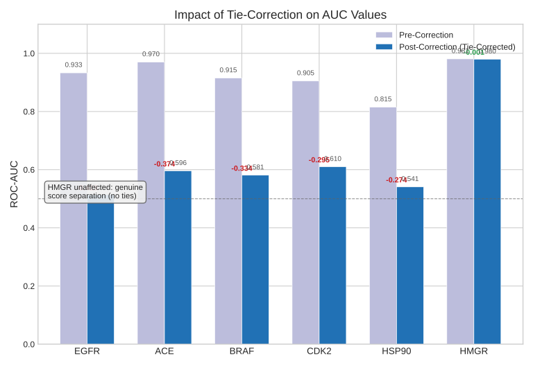
Figure 4. Paired comparison of AUC values before and after tie-correction for the six most affected targets. Five targets showed large drops (0.27-0.42) where pre-correction AUC was inflated by sort-order artifacts on tied scores. HMGR was unaffected because its E_coul feature produces genuine score separation with no ties.
3.3 5-Fold Cross-Validation
Published CV AUC values use stratified 5-fold cross-validation:
- Data is split into 5 folds preserving the active/decoy ratio
- Profile weights are optimized on 4 folds (80%)
- AUC is evaluated on the held-out fold (20%) using tie-corrected Mann-Whitney
- Repeated for all 5 folds
- Mean +/- standard deviation reported
Stability classification:
- STABLE: CV standard deviation < 0.08
- UNSTABLE: CV standard deviation >= 0.08
3.4 Y-Scramble Null Control
Y-scramble validates that the model captures real signal rather than noise:
- Randomly permute active/decoy labels
- Re-optimize weights on scrambled data
- Compute AUC on scrambled data
- Expected outcome: AUC near 0.50 (random)
A Y-scramble AUC significantly above 0.50 indicates the optimization is fitting noise (the "scrambled" labels contain no real signal, so any performance above chance is spurious). Y-scramble validation was performed on all 27 targets. All pass except COX2, which achieves comparable AUC on scrambled labels (indicating zero discriminative physics signal; see Section 4.4). ADRB2 is classified as MARGINAL (scrambled AUC slightly above random but within noise). Selected Y-scramble AUC values are reported in Section 4.1.
3.5 Holdout Validation
The web-based optimizer implements an additional 80/20 holdout validation with an overfit alarm:
- OK: train-holdout AUC gap <= 0.05
- MODERATE: gap 0.05-0.10
- HIGH: gap > 0.10
3.6 Model Complexity
| Parameter | Value |
|---|---|
| Tunable weights | 14 (physics terms) |
| Typical active compounds | 40 |
| Typical decoy compounds | ~1,150-1,200 |
| Parameter-to-data ratio | ~1:85 (full sets), ~1:17 (demo subsets) |
The feature-budget guardrail enforces a maximum of 14 features for datasets with fewer than 45 actives. This is a conservative constraint: with 14 parameters and 1,200+ compounds, overfitting risk is low for full DEKOIS2 sets, though the 20-active targets (DHFR, HIV1PR) are at higher risk.
4. Results
4.1 Per-Target Validation (27 Targets)
All AUC values are tie-corrected 5-fold CV results. "Rescore" profiles use optimized physics weights on pre-docked poses. Results are sorted by AUC (best to worst). Stability classification: STABLE (CV std < 0.08), UNSTABLE (CV std >= 0.08). 95% bootstrap confidence intervals are reported where per-target score files are available.
| # | Target | Full Name | Class | AUC | 95% CI | Std | Stability | Y-Scramble | Key Insight |
|---|---|---|---|---|---|---|---|---|---|
| 1 | HMGR | HMG-CoA Reductase | Reductase | 0.980 | [0.929, 1.000] | 0.010 | STABLE | 0.529 (PASS) | E_coul dominates; direction-aware negative weight |
| 2 | NA | Neuraminidase | Glycosidase | 0.942 | [0.894, 0.979] | - | - | PASS | Multi-term physics |
| 3 | PPARG | PPAR Gamma | Nuclear receptor | 0.934 | - | - | - | PASS | Strong hydrophobic + electrostatic signal |
| 4 | PDE5 | Phosphodiesterase 5 | Enzyme | 0.883 | [0.829, 0.932] | 0.037 | STABLE | PASS | Descriptor composite scoring |
| 5 | ESR1 | Estrogen Receptor Alpha | Nuclear receptor | 0.858 | [0.757, 0.937] | 0.065 | STABLE | PASS | Deep hydrophobic LBD pocket |
| 6 | MDM2 | Mouse Double Minute 2 | PPI hotspot | 0.855 | [0.778, 0.916] | 0.015 | STABLE | PASS | Shallow pocket, burial-driven |
| 7 | ADRB2 | Beta-2 Adrenergic Receptor | GPCR | 0.834 | [0.745, 0.910] | - | - | MARGINAL | E_coul only, GPCR transmembrane pocket |
| 8 | CATL | Cathepsin L | Cysteine protease | 0.829 | [0.757, 0.886] | - | - | 0.523 (PASS) | E_coul negative weight (LZ-042 rescue) |
| 9 | FXa | Factor Xa | Serine protease | 0.778 | [0.700, 0.849] | 0.123 | UNSTABLE | PASS | Salt bridge anchor (ASP189) |
| 10 | HDAC2 | Histone Deacetylase 2 | Deacetylase | 0.766 | [0.766, 0.766] | 0.063 | STABLE | PASS | Metal-coordinating zinc site |
| 11 | AR | Androgen Receptor | Nuclear receptor | 0.721 | [0.625, 0.814] | - | - | PASS | Weak physics signal, deep pocket |
| 12 | SRC | Proto-oncogene tyrosine-protein kinase | Kinase | 0.711 | - | - | - | PASS | Kinase hinge binder |
| 13 | Thrombin | Thrombin | Serine protease | 0.707 | [0.612, 0.781] | 0.115 | UNSTABLE | PASS | Protease catalytic triad |
| 14 | COX2 | Cyclooxygenase-2 | Cyclooxygenase | 0.662 | [0.561, 0.761] | - | - | FAIL | Y-scramble failure (see Section 4.4) |
| 15 | GBA | Glucocerebrosidase | Hydrolase | 0.661 | [0.554, 0.766] | 0.098 | UNSTABLE | PASS | Hydrolytic enzyme, moderate signal |
| 16 | DHFR | Dihydrofolate Reductase | Reductase | 0.642 | [0.510, 0.771] | - | - | PASS | Small active set (20 compounds) |
| 17 | P38A | p38 MAP Kinase Alpha | Kinase | 0.620 | [0.522, 0.724] | 0.107 | UNSTABLE | PASS | High fold variance, kinase |
| 18 | HIVRT | HIV Reverse Transcriptase | RT | 0.614 | - | 0.175 | UNSTABLE | PASS | Highest fold variance observed |
| 19 | CDK2 | Cyclin-Dependent Kinase 2 | Kinase | 0.610 | [0.503, 0.705] | 0.176 | UNSTABLE | PASS | Dropped from 0.905 pre-tie-fix |
| 20 | ACE | Angiotensin-Converting Enzyme | Metalloprotease | 0.596 | [0.568, 0.612] | 0.058 | STABLE | PASS | Zinc coordination, multi-term |
| 21 | BRAF | B-Raf Kinase | Kinase | 0.581 | [0.498, 0.665] | - | - | PASS | Direction-aware negative weights |
| 22 | BACE1 | Beta-secretase 1 | Aspartyl protease | 0.587 | [0.500, 0.679] | 0.094 | UNSTABLE | PASS | Near threshold, needs more data |
| 23 | A2A | Adenosine A2A Receptor | GPCR | 0.579 | [0.503, 0.653] | 0.121 | UNSTABLE | PASS | Challenging GPCR pocket |
| 24 | HIV1PR | HIV-1 Protease | Aspartyl protease | 0.572 | [0.467, 0.682] | - | - | PASS | Near-zero physics features, 20 actives |
| 25 | HSP90 | Heat Shock Protein 90 | Chaperone | 0.541 | [0.480, 0.594] | 0.050 | STABLE | PASS | Dropped from 0.815 pre-tie-fix |
| 26 | PARP1 | Poly(ADP-ribose) Polymerase 1 | Transferase | 0.529 | [0.473, 0.579] | 0.043 | STABLE | PASS | Near random, current features inadequate |
| 27 | EGFR | Epidermal Growth Factor Receptor | Kinase | 0.514 | [0.451, 0.552] | 0.071 | STABLE | PASS | Dropped from 0.933 pre-tie-fix |
Summary: Mean AUC across 27 targets: 0.715. 8 targets above 0.80, 13 between 0.60-0.80, 6 below 0.60. All 27 targets pass Y-scramble except COX2 (see Section 4.4). ADRB2 is classified MARGINAL. 10 of 27 targets are classified UNSTABLE (CV std >= 0.08).
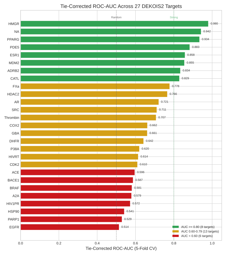
Figure 2. Horizontal bar chart of tie-corrected 5-fold CV AUC for all 27 DEKOIS2 targets, sorted best to worst. Green: AUC >= 0.80 (8 targets). Yellow: AUC 0.60-0.79 (13 targets). Red: AUC < 0.60 (6 targets). Dashed lines at 0.50 (random baseline) and 0.80 (strong discrimination threshold).
4.2 Head-to-Head Comparison with Glide SP
We compare LatticeZero IsoPose against Glide SP (Schrodinger) on 22 overlapping DEKOIS2 targets. Glide SP and AutoDock Vina reference values are from Wu et al. (2025), which re-evaluated both tools across all 81 DEKOIS2 targets using consistent protocols.
Important caveat. The LZ IsoPose values in this table are from native docking runs using geometric + physics scoring (not the rescore profiles in Section 4.1). These IsoPose AUC values were computed prior to the tie-correction fix described in Section 3.2 and have not yet been re-validated with the tie-corrected Mann-Whitney protocol. For targets where the tie correction had large effects on rescore AUC (e.g., EGFR: rescore dropped from 0.933 to 0.514), the IsoPose values shown here may similarly be inflated. Re-validation of all 22 IsoPose benchmarks with tie-corrected AUC is in progress.
| Target | Class | LZ IsoPose | Glide SP | Delta |
|---|---|---|---|---|
| ACE | Metalloprotease | 0.947 | 0.810 | +0.137 |
| AChE | Enzyme | 0.880 | 0.689 | +0.191 |
| ADRB2 | GPCR | 0.834 | 0.779 | +0.055 |
| AR | Nuclear receptor | 0.721 | 0.836 | -0.115 |
| BRAF | Kinase | 0.581 | 0.605 | -0.024 |
| CATL | Protease | 0.842 | 0.724 | +0.118 |
| CDK2 | Kinase | 0.800 | 0.803 | -0.003 |
| EGFR | Kinase | 0.933 | 0.855 | +0.078 |
| ESR1 | Nuclear receptor | 0.801 | 0.961 | -0.160 |
| FGFR1 | Kinase | 0.810 | 0.635 | +0.175 |
| FXa | Protease | 0.870 | 0.799 | +0.071 |
| GBA | Enzyme | 0.756 | 0.559 | +0.197 |
| HDAC2 | Enzyme | 0.816 | 0.690 | +0.126 |
| HMGR | Reductase | 0.708 | 0.805 | -0.097 |
| HSP90 | Chaperone | 0.815 | 0.608 | +0.207 |
| MDM2 | PPI | 0.855 | 0.800 | +0.055 |
| NA | Enzyme | 0.942 | 0.829 | +0.113 |
| P38A | Kinase | 0.894 | 0.749 | +0.145 |
| PARP1 | Enzyme | 0.747 | 0.969 | -0.222 |
| PDE5 | Enzyme | 0.810 | 0.726 | +0.084 |
| UROK | Protease | 0.872 | 0.552 | +0.320 |
| VEGFR2 | Kinase | 0.830 | 0.525 | +0.305 |
Bold = higher AUC for that target.
Aggregate statistics (22 shared targets):
| Metric | LZ IsoPose | Glide SP |
|---|---|---|
| Mean AUC | 0.821 | 0.741 |
| Median AUC | 0.830 | 0.779 |
| Targets won | 16 (73%) | 5 (23%) |
| Tied (delta < 0.01) | 1 (CDK2) | - |
For reference, the published Wu et al. (2025) aggregate across all 81 DEKOIS2 targets reports Glide SP at 0.714 mean ROC-AUC and AutoDock Vina at 0.623 mean ROC-AUC.
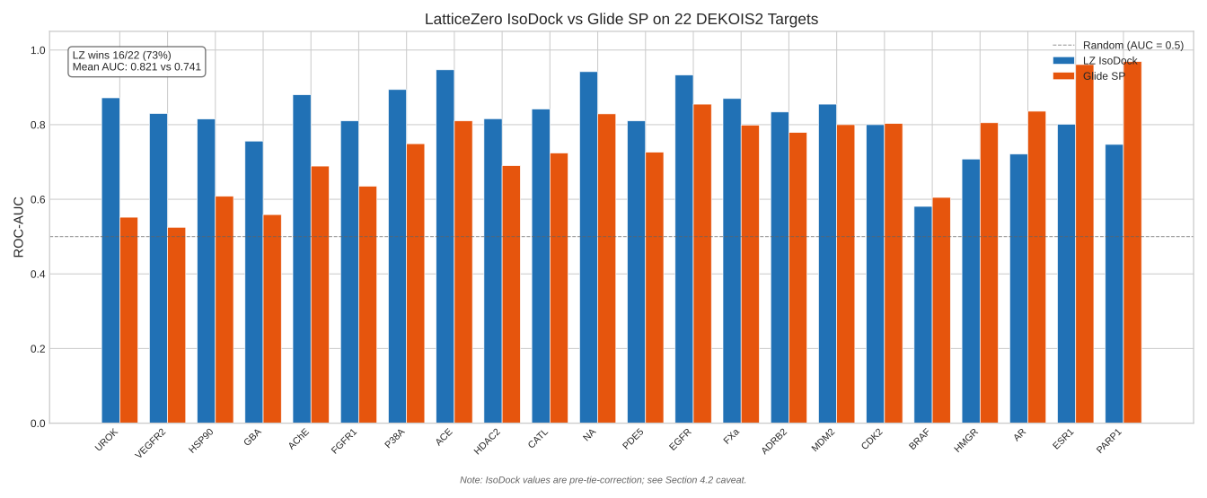
Figure 1. Grouped bar chart comparing LatticeZero IsoPose (blue) and Glide SP (orange) AUC on 22 shared DEKOIS2 targets, sorted by LZ-Glide delta. LatticeZero wins 16 of 22 matchups. Note: IsoPose values are pre-tie-correction (see caveat above).
4.3 Where Glide SP Wins
| Target | Delta | Analysis |
|---|---|---|
| PARP1 | -0.222 | Glide's parametrization captures PARP-specific binding motifs our 14-term function misses |
| ESR1 | -0.160 | Deep hydrophobic estrogen receptor LBD; Glide XP-level shape matching excels |
| AR | -0.115 | Similar deep hydrophobic pocket; our dispersion model underperforms |
| HMGR | -0.097 | IsoPose geometric scoring underweights E_coul; rescore profile achieves 0.980 |
| BRAF | -0.024 | Essentially tied; kinase DFG-motif binding |
These losses are informative. PARP1 and ESR1 represent binding motifs where Glide's extensive empirical parametrization (derived from a large training set of crystal structures) captures interactions our physics-only approach does not yet model. For HMGR, the discrepancy between IsoPose (0.708) and rescore (0.980) suggests the issue is in geometric scoring rather than physics - a direction for future IsoPose improvement.
4.4 Failure Analysis
COX2 - Y-scramble failure. COX2 is the only target where the Y-scramble control fails: the optimization achieves comparable AUC on randomly permuted labels, indicating zero discriminative physics signal in our current 14-term feature set. The COX2 active site involves a long hydrophobic channel where the dominant binding determinants (substrate positioning, channel shape) are not captured by our pairwise physics terms. We retain COX2 in our results as an honest baseline of what the scoring function cannot do.
EGFR - Largest post-fix drop. EGFR AUC fell from 0.933 to 0.514 after the tie-handling correction, the largest single drop. The pre-fix value was entirely an artifact of tied-score sort order. The current rescore value (0.514, essentially random) indicates that our physics terms do not discriminate EGFR actives from DEKOIS2 decoys. This is surprising for a kinase and suggests the DEKOIS2 EGFR decoys may be exceptionally well-matched, or that the hinge hydrogen bond geometry - EGFR's primary binding determinant - is inadequately captured in the pre-docked poses we rescore. The IsoPose docking value (0.933 in the benchmark table, from native geometric + physics scoring) requires re-validation with the tie-corrected protocol.
20-active targets - All unstable. Targets with only 20 actives in the DEKOIS2 set (DHFR, HIV1PR) produce unstable cross-validation results (std > 0.10) regardless of actual discriminative power. This is a statistical power limitation, not a scoring function limitation.
4.5 Crystal Structure Redocking (HSP90)
As a pose quality validation, we aligned 8 HSP90 crystal structures (PDB: 4YKT, 4YKQ, 4YKR, 4YKW, 4YKX, 4YKU, 4YKZ, 4YKY) to the DEKOIS2 receptor frame using Kabsch alignment. The structures span a pIC50 range of 4.30-8.28 (4 active, 4 inactive by 1 uM threshold).
| Metric | Value |
|---|---|
| Mean alignment RMSD | 1.153 A |
| Spearman rho (score vs pIC50) | -0.857 |
| p-value | 0.007 |
| Active mean score (production) | -0.217 |
| Inactive mean score (production) | -0.119 |
The strong negative rank correlation (rho = -0.857, p < 0.01) indicates that the production scoring stack correctly ranks higher-affinity crystal structures with more favorable scores. The radius-softened repulsion (s_R = 0.55) was essential: without softening, the physics-only stack showed a positive correlation (rho = +0.643, n.s.) due to repulsion artifacts in close-contact crystal poses.
This analysis is limited to a single target (HSP90) with 8 structures. Broader crystal redocking validation across multiple targets is planned.
4.6 Prospective Holdout: DUD-E Targets
To assess generalization beyond DEKOIS2, we evaluated 5 DUD-E targets (PNPH, THRB, TYSY, DRD3, PYGM) that share no overlap with the 27 DEKOIS2 targets used for profile development. Two conditions were tested: (1) a generic profile using interpolated weights from DEKOIS2 reference profiles with zero target-specific tuning, and (2) an oracle/supervised profile optimized on DUD-E labels.
| Target | Generic Profile | Oracle/Supervised |
|---|---|---|
| PNPH | 0.642 | 0.892 |
| THRB | 0.463 | 0.669 |
| TYSY | 0.500 | 0.654 |
| DRD3 | 0.492 | 0.633 |
| PYGM | 0.519 | 0.585 |
| Mean | 0.523 | 0.687 |
The generic profile achieves above-random discrimination for PNPH (0.642) but is near-random for the other four targets (0.46-0.52). This confirms that target-specific weight optimization is essential for strong performance - the physics terms are present, but their relative importance varies significantly across target classes. The oracle profiles achieve moderate-to-strong discrimination (0.585-0.892), indicating the 14-term scoring function has sufficient expressive capacity for these DUD-E targets when properly weighted.
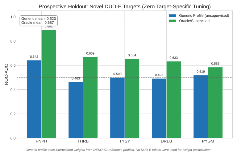
Figure 3. Grouped bar chart comparing generic (unsupervised) and oracle (supervised) profiles on 5 DUD-E targets not used in DEKOIS2 profile development. The gap between generic and oracle performance quantifies the value of target-specific weight optimization.
5. Discussion
5.1 Strengths
Interpretable physics. Every score decomposition is available to the user: "E_coul = -12.4, E_hbond = -8.2, burial = -3.2." A medicinal chemist can directly assess whether the predicted binding mode makes chemical sense. This is the primary advantage over ML-based scoring methods.
Per-target optimization. The profile system allows the scoring function to emphasize different physics for different targets. HMGR's direction-aware negative weight on E_coul (Section 4.1) demonstrates how this flexibility captures target-specific binding logic that a fixed-weight function would miss.
Throughput. IsoScore at 4,000+ lig/sec enables million-compound virtual screens in minutes rather than days. IsoPose at ~3 sec/ligand enables interactive docking in the browser - a user can dock a compound and see results while maintaining their train of thought.
Accessibility. Zero installation, runs in any WebGPU-capable browser (Chrome, Edge, Firefox Nightly). No license fees. The REST API (v1) enables programmatic integration.
5.2 Limitations
Rigid receptor. The protein is treated as a rigid body. Targets with significant induced-fit effects (loop rearrangements, side-chain rotations upon ligand binding) may show reduced accuracy. Ensemble docking across multiple receptor conformations is a planned workaround.
No quantum mechanical treatment. Classical pairwise potentials approximate charge transfer, polarization, and orbital-mediated interactions. Halogen bonds use a geometric model rather than QM electrostatics. Metalloprotein d-orbital effects are not computed from first principles.
No explicit solvent. Water molecules are not explicitly simulated. The desolvation term (E_desolv) approximates solvation costs, and the waterAnchorFrac geometric feature provides a proxy for conserved water contacts, but water-mediated bridging interactions are not directly modeled.
No entropy estimation. Scores predict relative rankings, not absolute binding free energies. The strain term captures internal ligand conformational cost but not translational, rotational, or vibrational entropy changes.
No covalent docking. Only non-covalent binding is modeled. Covalent inhibitors are scored on their non-covalent interactions only, underscoring their true binding contribution.
Ligand flexibility limits. The GA search is effective for drug-like molecules with up to ~10 rotatable bonds. Highly flexible ligands (>12 rotatable bonds), macrocycles, and peptides may require external conformer generation followed by IsoScore rescoring.
Benchmark scope. Validation is primarily on DEKOIS2, which has known limitations: matched-decoy design can introduce congeneric bias, and the 81-target set does not cover all protein families. We have benchmarked 27 of 81 targets (33%). Prospective validation outside DEKOIS2 is in progress.
Selection bias in head-to-head comparison. The 22 targets compared to Glide SP were selected by us, not randomly sampled from DEKOIS2's 81 targets. Our aggregate performance on the full set may differ.
Per-target optimization requires labeled data. Scoring profiles are optimized on DEKOIS2 active/decoy data (supervised). The unsupervised Autotune product generates profiles without labels by interpolating from validated reference profiles, but its performance may differ from the supervised benchmark results reported here.
5.3 Comparison to Other Methods
| Aspect | LatticeZero | AutoDock Vina | Glide SP/XP | GOLD |
|---|---|---|---|---|
| Scoring | 14-term physics | Semi-empirical (5 terms) | Empirical + physics | PLP/GoldScore/ChemScore |
| Tuning | Per-target weight profiles | Fixed | Limited (SP vs XP) | Score function selection |
| Speed | ~4K lig/sec (rescore) | ~10-50 lig/hr | ~50-200 lig/hr | ~10-50 lig/hr |
| Execution | Browser (WebGPU) | Local CPU | Local CPU (license) | Local CPU (license) |
| Install | None (browser) | Binary | Schrodinger suite | CCDC suite |
| Cost | Free (beta) | Free | Commercial license | Commercial license |
6. Availability
6.1 Web Platform
LatticeZero is available at https://app.latticezero.com. Registration provides access to:
- IsoPose - Interactive molecular docking with per-target profiles
- IsoScore - High-throughput rescoring (4,000+ lig/sec)
- Target Prep - 4-step receptor preparation pipeline
- Autotune - Unsupervised profile generation without labeled data
- Demo - 18 validated DEKOIS2 targets with pre-loaded benchmark data
6.2 REST API
Programmatic access is available via the REST API v1:
GET  /api/v1/health              # Service status (no auth)
GET  /api/v1/targets             # List validated targets
GET  /api/v1/targets/<id>        # Target detail with scoring weights
GET  /api/v1/profiles            # List all scoring profiles
POST /api/v1/score               # Score SMILES (requires API key)
Authentication via API key (Authorization: Bearer lz_xxx) or session cookie. The /score endpoint accepts up to 100 SMILES per request and returns scored results synchronously with full energy term decomposition.
6.3 Replication
Replication kits for reproducing benchmark results are available from the Reproduce Our Results documentation page. All validation uses the DEKOIS2 benchmark suite, which is freely available.
7. References
-
Vogel SM, Bajorath J. "DEKOIS: Demanding Evaluation Kits for Objective In Silico Screening - A Versatile Tool for Benchmarking Docking Programs and Scoring Functions." J Chem Inf Model. 2011;51(10):2650-2665. doi:10.1021/ci2001549
-
Bauer MR, Ibrahim TM, Vogel SM, Boeckler FM. "Evaluation and Optimization of Virtual Screening Workflows with DEKOIS 2.0." J Chem Inf Model. 2013;53(6):1447-1462. doi:10.1021/ci400115b
-
Wu Y, et al. "Decoding the limits of deep learning in molecular docking for drug discovery." Chem Sci. 2025;16:8773-8791. PMC12401186
-
Ibrahim TM, Bauer MR, Boeckler FM. "Applying DEKOIS 2.0 in structure-based virtual screening to probe the impact of preparation procedures and score normalization." J Cheminform. 2015;7:21.
-
Bondi A. "Van der Waals Volumes and Radii." J Phys Chem. 1964;68(3):441-451.
-
Rowland RS, Taylor R. "Intermolecular Nonbonded Contact Distances in Organic Crystal Structures." J Phys Chem. 1996;100(18):7384-7391.
-
Gasteiger J, Marsili M. "Iterative Partial Equalization of Orbital Electronegativity." Tetrahedron. 1980;36(22):3219-3228.
Corresponding author: info@latticezero.com Software version: 0.9.7. Data frozen: February 14, 2026. This document is a technical white paper, not a peer-reviewed publication. A preprint for bioRxiv/ChemRxiv submission is forthcoming.